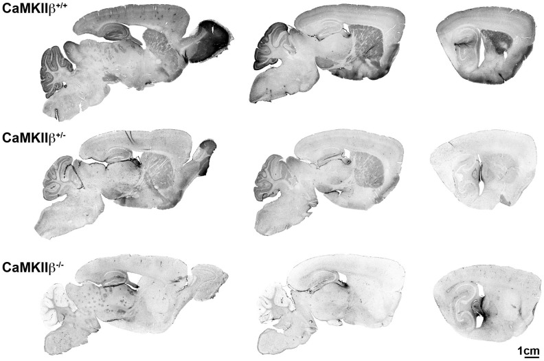
Figure 2. CaMKIIβ immunohistochemistry.
Representative images of the CaMKIIβ IHC staining for one mouse from each genotype is shown at approximately 1 mm lateral intervals from midline. In the WT (+/+) mouse, CaMKIIβ staining is highest in the olfactory bulb, but is also apparent in the cerebellum, cortex, hippocampus, striatum, and substantia nigra. The heterozygous mice show an intermediate level of staining. The KO mice show a lack of CaMKIIβ staining throughout the brain.

