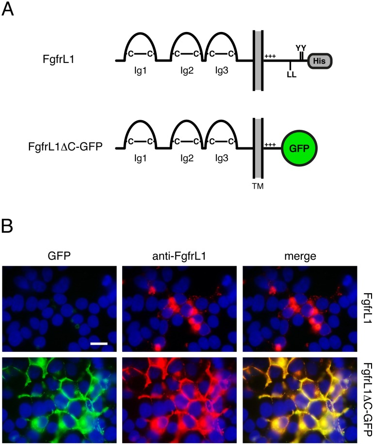
Figure 1. FgfrL1ΔC-GFP protein remains at the plasma membrane.
A) Schematic drawing of full-length FgfrL1 and of truncated FgfrL1ΔC-GFP. The three Ig domains with disulfide bridges (C–C), transmembrane domain (TM), positively charged juxtamembrane region (+++), dileucine motif (LL), tandem tyrosine-based motif (YY), histidine-rich region (His) and GFP moiety are indicated. B) HEK293 cells were transfected with constructs coding for full-length FgfrL1 and for FgfrL1ΔC-GFP as indicated. After one day, the cells were fixed and stained with a monoclonal antibody against FgfrL1. Full-length FgfrL1 was localized primarily to intracellular structures, whereas the mutant protein was found mainly at the cell membrane. The signal from GFP (green) and from the antibody (red) colocalized as demonstrated by superimposition of the two panels. Cell nuclei were stained with DAPI (blue). Bar = 20 µm.

