
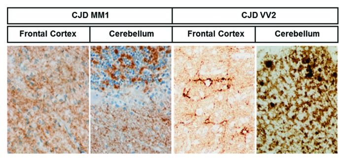
Figure 3. PrPsc deposition in sCJD brain samples. PrPsc immunohistochemistry shows differential PrP deposition in frontal cortex and cerebellum in sCJD MM1 and sCJD VV2 cases. Sections were pre-incubated with PK prior to PrPsc immunohistochemistry. Synaptic pattern of PrPsc deposition in the frontal cortex and molecular layer of the cerebellum characterizes MM1, whereas synaptic and plaque-like PrPsc deposition, mainly in the cerebellum, characterizes VV2.