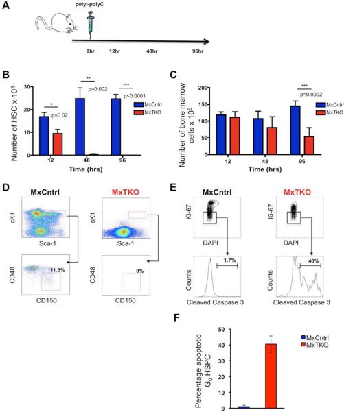
Figure 2. Apoptosis of Hematopoietic Stem/Progenitor Cells Following Shutdown of D-cyclins.
(A) Outline of the experimental design. Mx-Cre+;D1F/FD2−/−D3F/F (MxTKO) and control Mx-Cre+;D1F/+D2+/−D3F/+(MxCntrl) animals received a single injection of pI-pC (to delete D-cyclins in MxTKO mice). Animals were sacrificed after 12, 48 and 96 hours, and hematopoietic stem cells (HSC) were stained and quantified by FACS.
(B and C) Mean number of HSC (B), and mean number of total bone marrow cells (C), per animal (including femurs, tibias, Iliac crests and humerus) at the indicated time-points after ablation of D-cyclins.
(D) Representative FACS analysis of HSC (Linlow, Kit+, Sca1+, CD48−, CD150+) from bone marrows collected at 96 hours after deletion of D-cyclins. Note that essentially no HSC were detected in MxTKO animals.
(E) 24-48 hours after ablation of D-cyclins, hematopoietic stem/progenitor cells (HSPC) were stained with DAPI (4’,6-diamidino-2-phenylindole), Ki67 and cleaved caspase 3 (to mark apoptotic cells). HSPC then were gated on G0 cells (Wilson et al., 2008) and analyzed for cleaved caspase 3 staining.
(F) The mean percentage of caspase 3-positive cells among quiescent HSPC cells, gated as above, in MxCntrl and MxTKO animals.
In (B), (C) and (F), error bars denote SD.
See also Figure S2.

