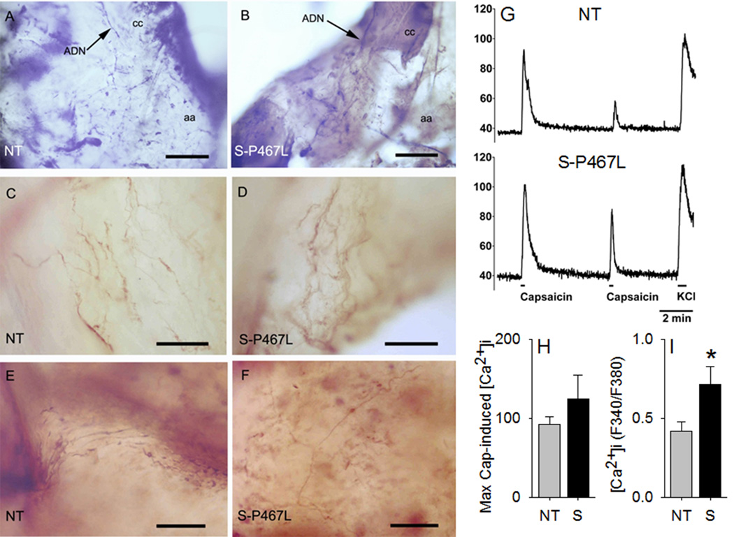
Figure 7. Neurovascular Coupling and Function of Trpv1 Channels in Nodose Ganglia Neurons.
Fibers and terminals of the aortic depressor nerve (ADN) on the aortic arch (aa) immunochemically stained with an antibody against Trpv1 in non-transgenic (NT) and S-P467 transgenic mice. A) Low power micrograph from a whole mount preparation from a NT animal showing the ADN innervating the region of the aortic arch at the junction with the left common carotid artery (cc). B) Similar preparation as in A from a S-P467L mouse. Note the similar appearance of the ADN and its arborizations in both mice. C–D) Complex endings of Trpv1 positive ADN axons in the vicinity of the aortic arch-left carotid junction. E–F) Arborizations of thin Trpv1 positive axons on the anterior wall of the aortic arch. Scale bars in A and B=250μm; scale bars in C–F=25μm. G) Representative traces of Ca2+ imaging in response to two successive capsaicin applications (100 nM, 15 sec), followed by KCl application (50 mM, 30 sec) in NG neurons. H) Maximal capsaicin-induced [Ca2+]i responses normalized to KCl. I) Magnitude of Trpv1 desensitization, expressed as the ratio of 2nd vs 1st capsaicin-induced peak [Ca2+]i in the same neuron. N=38 for NT and N=58 for S-P467L. *, P<0.05 vs NT.

