
Figure 3.
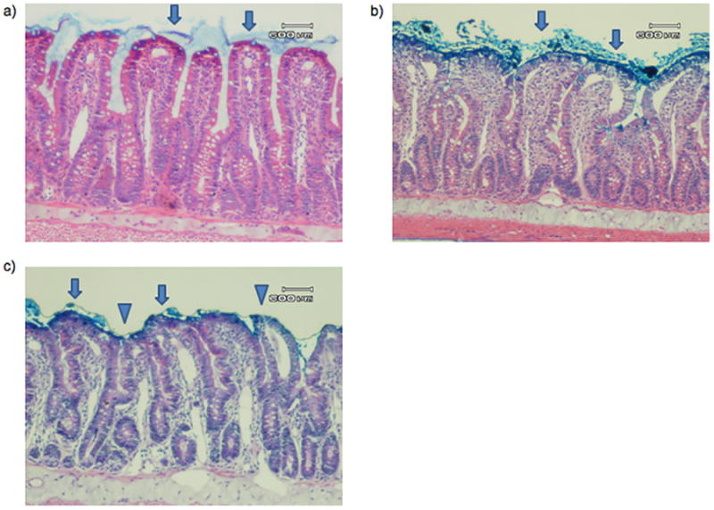
Representative photomicrograph of a) SP, b) PDD, and c) AP histology. In both of the sham pancreatitis groups, including a) sham pancreatitis and b) pancreatic duct diversion sham pancreatitis, villi are covered with an intact mucus layer (arrow). In the acute pancreatitis group, some villi are covered with an intact mucus layer (arrow), but the majority of villi demonstrate mucus sloughing (triangle). There is no significant difference noted in injured villi between groups. A 600μm reference is provided for scale.