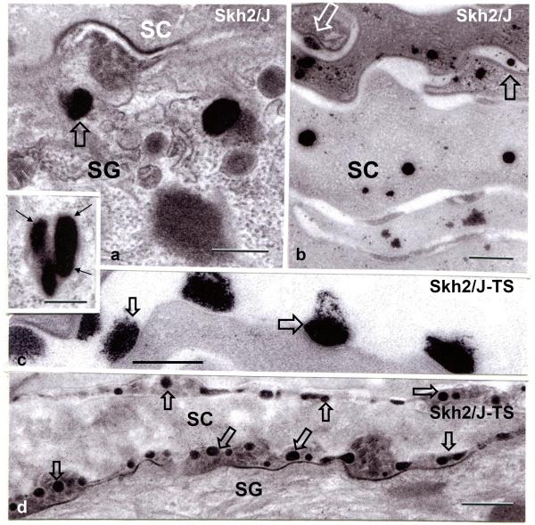
Figure 4. Pigment Granule Persistence, with Rapid Extrusion after Barrier Disruption in SKH2/J Mice.
A: Under basal conditions, large pigment granules persist into the SG and SC, where they largely disintegrate within the corneocyte cytosol (B). Under basal conditions, some granules appear to be extruded into the extracellular spaces (B, open arrows). A, insert: pigment granules appear enclosed within membrane bound organelles (likely phagolysosomes) in the outer nucleated cell layers of the SKH2/J epidermis. Shortly after acute barrier disruption by tape stripping (SKH2/J TS), pigment granules are extruded at the SG–SC interface, and also within the SC extracellular spaces in SKH2/J mice (C&D, open arrows). Osmium tetroxide post-fixation. Mag bars, A-C + insert: 0.25 μm, D: 0.5 μm.

