Fig. 1.
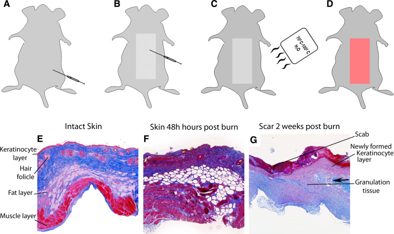
Experimental steps in the burn rodent model and histological images of C57/BL mice skin subjected to full-thickness burn (30 %TBSA). a The rodent is anesthetised with an intraperitoneal injection of Xylazine and Ketamine. b The area (dorsum) to be burned is shaved with a clipper to ensure an even burn. c The rodent is then placed in a flame-resistant mold with an opening exposing a pre-determined total body surface area to burn; the exposed area is then immersed in a 100 °C water bath for 8 s. d Lactated Ringer’s solution is then administered intraperitoneally for resuscitation; buprenorphine or other analgesia may be administered subcutaneously for pain control. Excised burned skin tissue specimens from burned mice (thickness = 5 μm) were harvested and then Masson’s trichrome staining performed. e Intact skin showing histological component of mouse non-burned skin. f Burned skin harvested from mouse 48 h post-burn. Note that the animal is presenting with complete destruction of skin, most obviously in the epidermal/dermal segments. g Animal at 2 weeks post-burn showing signs of wound healing: re-epithelialization (at wound edge), neovascularization, and formation of new granulation tissue. Arrows indicate wound edges or new granulation tissue formation. Collagen fibers in the dermis are stained in blue, epidermis and muscle stained in red

