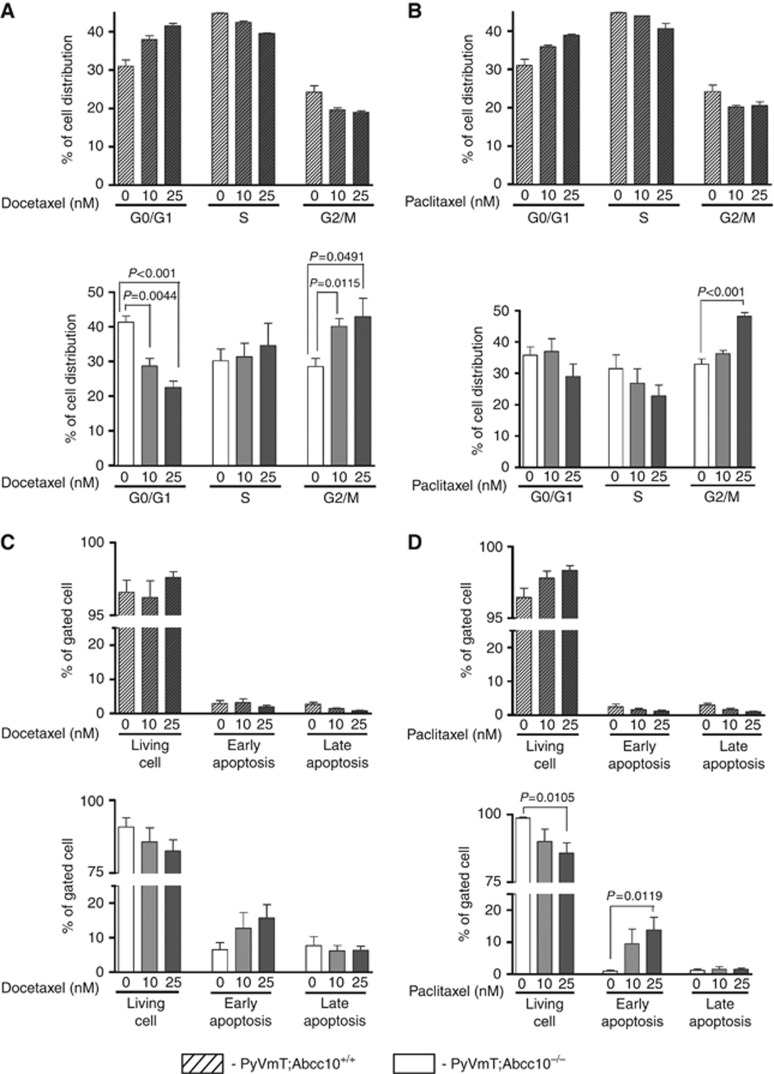
Figure 5.
Effects of docetaxel and paclitaxel on cell cycle progression and apoptosis induction in MMTV-PyVT;Abcc10+/+ and MMTV-PyVT; Abcc10−/− primary mammary tumours cells. After treatment with 0, 10, or 25 nM docetaxel or paclitaxel, cells were harvested for analysis of cell cycle distribution or apoptosis. Cell cycle distribution is shown after 48 h of docetaxel (A) or paclitaxel (B) treatment. Percentage of living cells, and early and late apoptosis are shown following 48 h of docetaxel (C) or paclitaxel (D) treatment. Shade, MMTV-PyVT;Abcc10+/+ cell lines; blank, MMTV-PyVT;Abcc10−/− cell lines. Error bars denote mean±s.e.m. Statistical significance was determined by two-sided Student's t-test.

