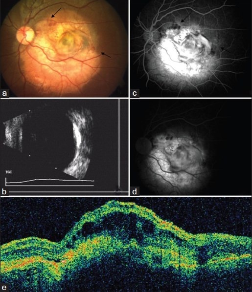
Figure 1.

Left eye, at presentation (a) Fundus photograph showing choroidal osteoma with choroidal neovascularization (CNV) and subretinal hemorrhage (arrows) (b) B-scan showing hyperechoic osteoma with orbital shadowing (c) Fundus fluorescein angiography (FFA) early phase showing hyperfluorescence of CNV and blocked fluorescence of hemorrhage (arrows) (d) FFA late phase showing late leakage of CNV (e) Optical coherence tomography (OCT) scan showing retinal pigment epithelium (RPE) disruption, overlying cystoid macular edema (CME) and minimal subretinal fluid (SRF) suggestive of active CNV
