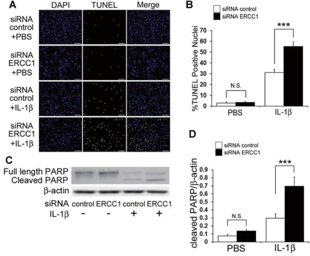
Figure 3.
Effect of ERCC1 inhibition on cell death in HC under IL-1β stimulation. (A) Representative images of TUNEL staining after inhibition of ERCC1 under IL-1β stimulation. HC were immunostained for TUNEL (green) and DAPI (blue). Scale bar = 100µm. (B) Quantification of %TUNEL positive cells was calculated as the percent of cells (DAPI) expressing TUNEL (green). Error bars indicate the SD. ***p <0.001. (C) Representative image of an immunoblot to measure the protein level of full length and cleaved PARP after inhibition of ERCC1 when stimulated with IL-1β. (D) Quantification of the expression ratios of cleaved PARP to β-actin was calculated. Error bars indicate the SD. ***p < 0.001, not significant (NS).

