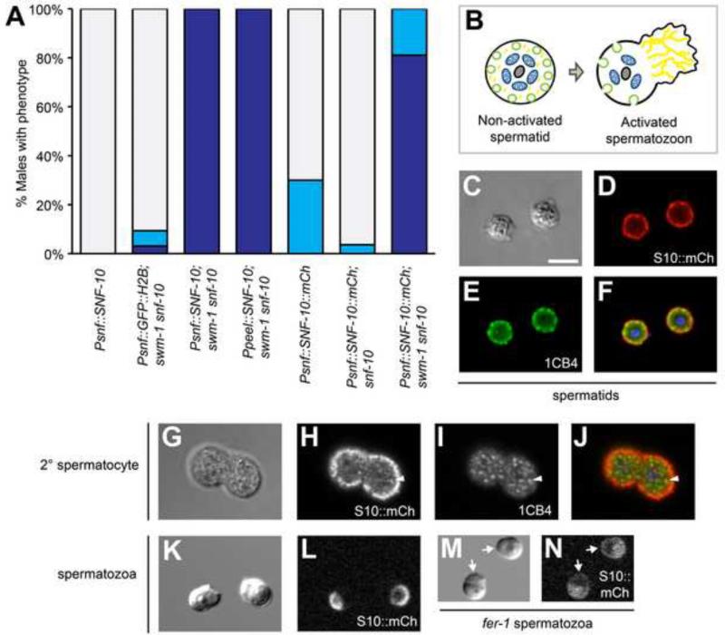
Fig. 3. snf-10 is expressed in and functions in sperm.
(A) Rescue of the sperm activation phenotype by snf-10 transgenes. Either a snf-10 or sperm-specific peel-1 promoter can confer rescue, as can expression of SNF-10::mCherry reporter protein. As in Fig. 1E, column shading indicates the percent of males containing only activated sperm (dark blue), a mixture of spermatids and activated sperm (light blue), or only non-activated sperm (grey); n, 30-55. (B) Schematic of spermatid and spermatozoon structure. Grey, chromatin; blue, mitochondria; green, MOs; yellow, MSP cytoskeleton. (C-J) In jnSi96[Psnf-10::SNF-10::mCh] spermatids, SNF-10::mCh and 1CB4 are both localized primarily to the cell periphery; some concentrations of mCh and 1CB4 are clearly distinct from one another. In 2° spermatocytes, most mCh is on the cell cortex and fails to colocalize with 1CB4, but some cytoplasmic puncta are visible. Arrowhead indicates a region of overlap between mCh and 1CB4. Images are fixed cells visualized by (C,G) DIC, (D,H) anti-RFP staining, (E,I) 1CB4 staining, and (F,J) a merge of anti-RFP, 1CB4, and DAPI. (K-N) SNF-10::mCh is polarized to the cell body plasma membrane in activated spermatozoa, but distributed throughout the plasma membrane in a fer-1 mutant. Live sperm were visualized by (K,M) DIC and (L,N) mCherry fluorescence. Arrows indicate SNF-10::mCh in fer-1 pseudopodia. (A-N) All strains were mutant for unc-119 and him-5, as well as snf-10(hc194) except as indicated in (A). (M,N) Worms were raised at 25°C, the non-permissive temperature for fer-1(hc1). Scale bar for all images, 5 microns.

