
Fig. 7.
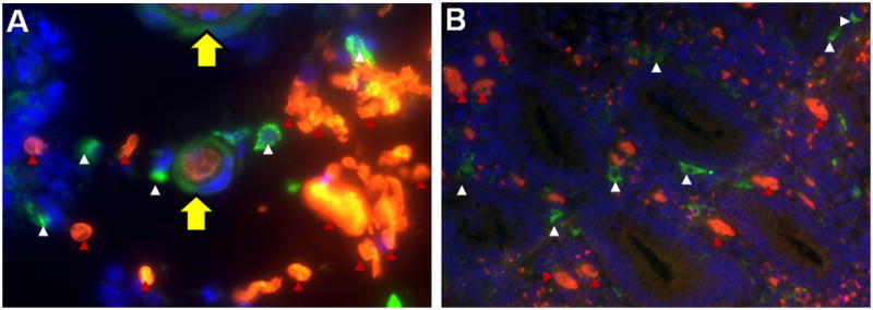
Double immunofluorescent staining for hemoglobin-α (red) and the macrophage marker CD163 (green) in paraffin-embedded sections from a patient with UC. Nuclei were counterstained with DAPI (blue): red arrows indicate extravasated erythrocytes and white arrows indicate macrophages. Pictures were taken at 60× (A) and 20× (B) magnification. A) Depicts macrophage erythrophagocytosis: a macrophage (yellow arrow) engulfed three red blood cells from extravasated erythrocytes (red arrow). B) Colonic mucosa with extravasated erythrocytes and macrophages in close vicinity to crypts.