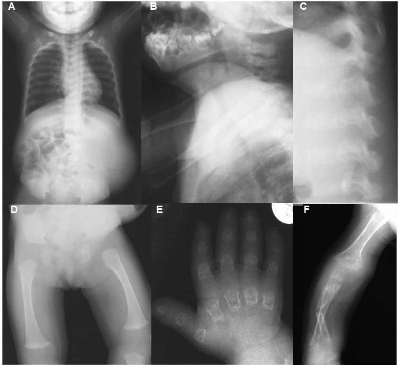
FIG. 2.
Radiographs of the patient at 21 months (A-C) and 7 years (D-F) of age. (A) Anterior-posterior (AP) image showing a small thorax and small, rounded pelvis. (B and C) Lateral view of neck and spine showing platyspondyly with under mineralization of the cervical (B) and lumbar (C) vertebrae. (D) AP view of the femur showing severe epiphyseal delay of the capital femoral epiphysis and flared metaphyses of the distal femur. (E) AP view of the hand showing characteristically short and irregular metacarpals and phalanges with severely reduced carpal ossification and osteoporosis. (F) AP view of an upper extremity showing a fractured radius and generalized undermineralization of the upper limb.

