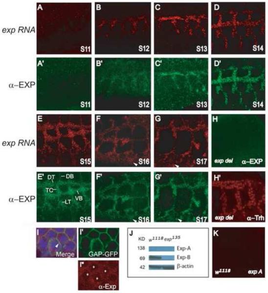
Fig. 3. Exp is localized throughout the cytoplasm of all tracheal cells.
The localization of exp RNA and Exp protein was analyzed by fluorescence in situ hybridization (FISH) to an exp RNA probe and followed by immunostaining with anti-Exp antisera using whole mount wild type embryos. The exp probe and the anti-Exp antisera recognize both isoforms. Sagittal views of tracheal segments 3-7 are shown, anterior is to the left and dorsal is up. Dorsal trunk (DT), transverse connective (TC), lateral trunk (LT), visceral branch (VB), and dorsal branch (DB) were labeled. (A-G) exp RNA is expressed throughout the trachea from stages 12-17, but not at stage 11. Additionally, exp RNA is also observed in epidermal cells at stage 16 and 17 (arrowheads). (A’-G’) The Exp protein has the exact same expression pattern as exp RNA. It is expressed in all tracheal cells from stages 12-17, but not at stage 11. The Exp protein is also observed in epidermal cells at later stages (arrowheads). To visualize intracellular localization of Exp, wild type embryos containing btl-Gal4 UAS-GAP-GFP were co-immunostained with anti-Exp (red in I, I”), anti-GFP that stains the cell membrane marker GAP-GFP (green in I, I’), and anti-Trh that stains tracheal nuclei (blue in I), shown in DT cells. Exp is clearly localized throughout the cytoplasm (red in I) and also partially overlaps with the membrane (yellow in I; arrowhead). (H) Exp protein is not detected in homozygous Df(2R)BSC879 embryos, which delete the entire exp gene locus and several other genes, even though tracheal cells are present (labeled by Trh (red) in H’). (J) The 69KD Exp-B isoform is detected in cell lysate from wild type but not exp135 mutant embryos by Western blot using anti-Exp antisera. However, the 139KD Exp-A is not detected in wild type. (K) exp-A RNA is not detected by FISH using an exp-A RNA probe that only recognizes the exp-A isoform.

