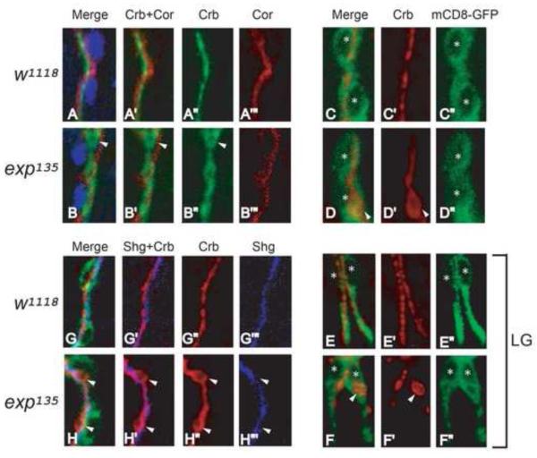
Fig. 5. Cysts are apical compartments within the cell boundary in unicellular and intracellular branches in exp mutants.
(A-B”’) Stage 15 w1118 and exp135 embryos were immunostained with the apical membrane marker Crb, basolateral membrane marker Cor, and tracheal nuclei marker Trh, to visualize apical-basal polarity in wild type and exp135 mutant embryos. Unicellular branch GBs were shown. Both Crb (green) and Cor (red) appear as single lines without overlapping in wild type embryos (A-A”’). Similarly, cysts outlined by Crb do not overlap with Cor in exp135 mutant embryos (B-B”’), indicating that cysts are apical compartments. (G-H”’) Stage 15 w1118 and exp135 embryos containing btl-Gal4 mCD8-GFP that outlines the cell boundary were immunostained for Crb and the AJ marker Shg. Shg (blue) labels a line of autocellular junctions along the apical membrane (red) in wild type GB (G-G”’). Similarly, Shg (blue) labels a line of autocellular junctions despite cyst (red) formation in exp135 embryos, suggesting that cysts are part of the extracellular lumen in unicellular branches (HH”’). (C-F”) Stage 15 w1118 and exp135 embryos containing btl-Gal4 mCD8-GFP were immunostained for Crb and the cell membrane marker GFP. In wild type embryos, Crb (red) appears as a line within the cell boundary marked by mCD8-GFP (green) in unicellular branch GB (C-C”) and intracellular branch LG (E-E”). Similarly in exp135 mutant embryos, cysts (white arrowheads) in GB (D-D”) and LG (F-F”) are also within the mCD8-GFP labeled cell boundary. Nuclei were labeled by asterisks.

