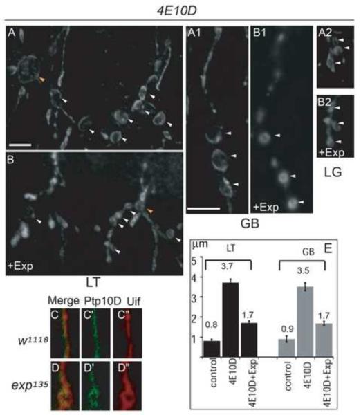
Fig. 7. Exp expression suppresses cyst phenotype in Ptp4EPtp10D double mutants, and Exp is not required for the distribution of Ptp10D.
Stage 16 embryos were stained with anti-Uif to label the tracheal apical membrane and imaged using confocal microscopy. LT in tracheal segments 5-6 were shown, GB in tracheal segment 6 was shown. (A-B2) Expression of HA-Exp-B in Ptp4EPtp10D embryos significantly reduced cyst size in LT (B), GB (B1) and LG (B2) compared to cysts in Ptp4EPtp10D embryos (A, A1, A2). In addition, TC/LT branch junction cyst size was also significantly reduced. White arrowheads point to LT and GB cysts and orange arrowheads point to TC/LT junction cysts. Quantification of LT and GB cysts is shown by the bar graph (E). Single GB stained with anti-Ptp10D (green) and the apical membrane marker Uif (red) in wild type (C-C”) and exp135 mutant embryos (D-D”) were shown. Ptp10D (C-C’) was localized at the apical membrane labeled with Uif (C”) in wild type embryos. Similarly, Ptp10D (D-D’) was also localized around cysts outlined with Uif (D”). The white line in B represents 10 μm in A and B. The white line in A1 represents 10 μm in the rest of the panels.

