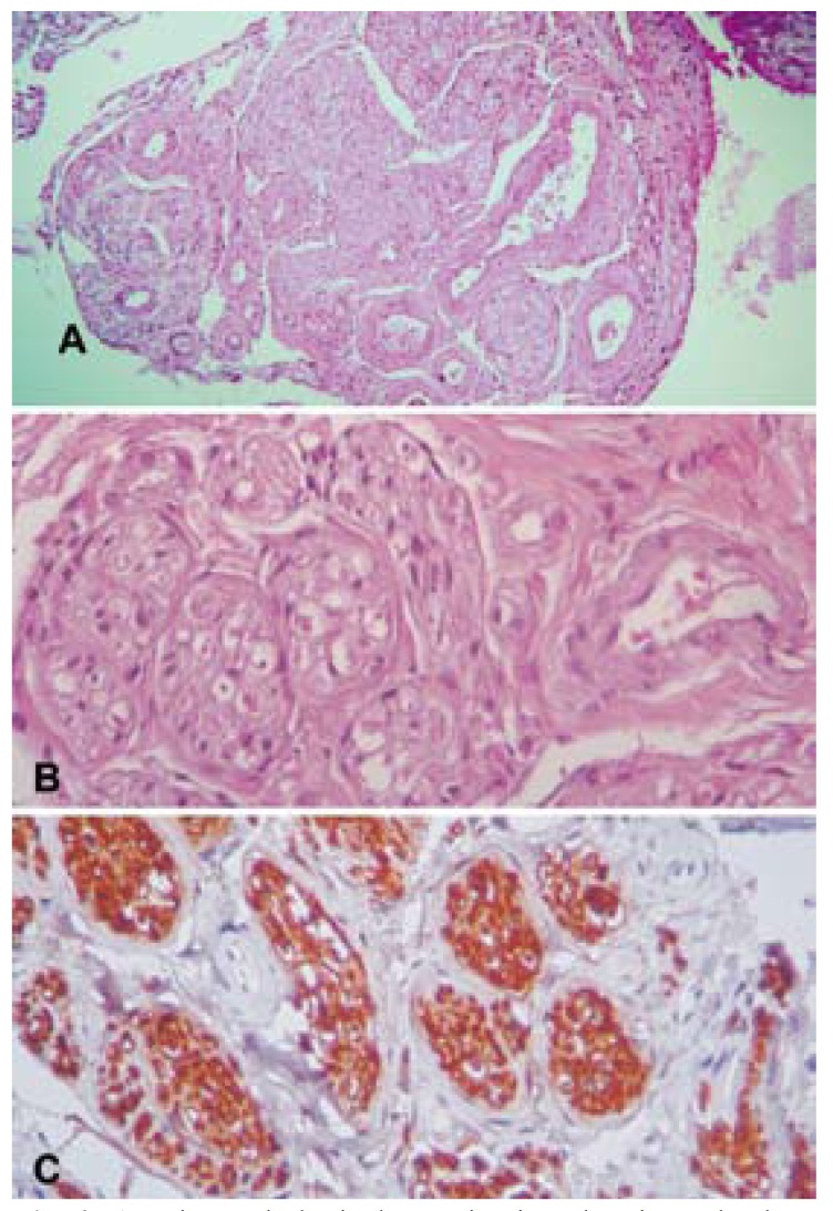
Figure 2.
A. Histopathological examination showing a haphazard, tortuous proliferation of nerve bundles within a vascularized fibrous connective tissue stroma. Hematoxylin and eosin stain, original magnification x10; B. Cross-sectioned nerve bundle within a vascularized fibrous connective tissue stroma. Hematoxylin and eosin stain, original magnification x40; C. Strong positivity of the bundles for the neural marker S-100. Immunohistochemical staining, original magnification x40.

