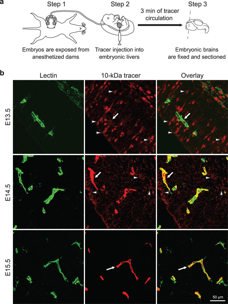
Figure 1. A novel tracer injection method reveals a temporal profile of functional BBB formation in the embryonic cortex.
a, In-utero embryonic liver tracer injection method - fenestrated liver vasculature allowed rapid tracer uptake into the embryonic circulation. b, 10-kDa dextran-tracer injection revealed a temporal profile of functional cortical BBB formation. Representative images of dorsal cortical plates from injected embryos after capillary labeling with lectin (Green: lectin, red: 10-kDa tracer). Upper panel, E13.5: Tracer leaked out of capillaries and was subsequently taken up by non-vascular parenchyma cells (arrowheads), with little tracer left inside capillaries (arrow). Middle panel, E14.5: Tracer was primarily restricted to capillaries (arrow), with diffused tracer detectable in the parenchyma (arrowheads). Lower panel, E15.5: Tracer was confined to capillaries (arrow). n=6 embryos (3 litters/age).

