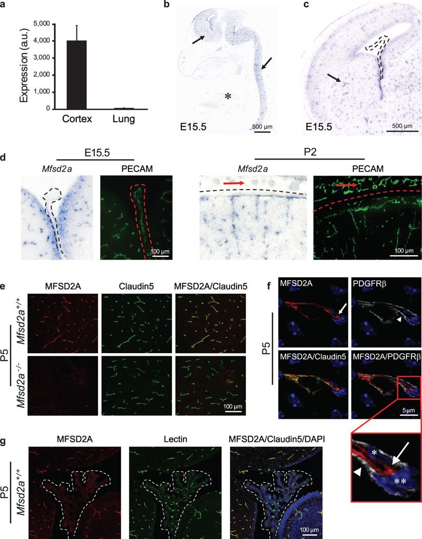
Figure 3. Mfsd2a is selectively expressed in BBB-containing CNS vasculature.
a, E13.5: Mfsd2a expression in cortical endothelium was ~80-fold higher than lung endothelium (microarray analysis, mean±s.d.). b-d, Specific Mfsd2a expression in BBB-containing CNS vasculature (Blue: Mfsd2a in situ hybridization, green: vessel staining (PECAM) adjacent sections). b, Mfsd2a expression in CNS vasculature (E15.5 sagittal view-brain and spinal cord, arrows), but not in non-CNS vasculature (asterisk). c, Mfsd2a expression in BBB vasculature (E15.5 cortex coronal view, e.g. striatum, arrow), but not in non-BBB CNS vasculature (choroid plexus, dotted line). d, High magnification coronal view of Mfsd2a expression in BBB-containing CNS vasculature but not in vasculature of the choroid plexus (left, dotted line), or outer meninges/skin (right, red arrows). e-g, Immunohistochemical staining of MFSD2A protein shows specific expression in CNS endothelial cells (Red: MFSD2A; green: Claudin5 or Lectin (endothelium); blue: DAPI (nuclei); gray: PDGFRβ (pericytes)). e, MFSD2A expression in the brain vasculature of wild-type mice (upper panel), but not of Mfsd2a−/− mice (lower panel). f, MFSD2A expression only in Claudin5-positive endothelial cells (arrow, endothelial nucleus–asterisk) but not in adjacent pericytes (arrow head, pericyte nucleus–double asterisk). g, Lack of MFSD2A expression in choroid plexus vasculature (fourth ventricle coronal view-dotted line), as opposed to the prominent MFSD2A expression in cerebellar vasculature. n=3 embryos (3 litters/age).

