
Fig. 4.
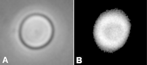
A red blood cell measured using the 1200 MHz transducer. (A) Optical and (B) Photoacoustic image of a single RBC fixed on the glass substrate. The contours the the RBC are clearly visible in both images. The width of the image is 15 μm.
Official websites use .gov
A
.gov website belongs to an official
government organization in the United States.
Secure .gov websites use HTTPS
A lock (
) or https:// means you've safely
connected to the .gov website. Share sensitive
information only on official, secure websites.
A red blood cell measured using the 1200 MHz transducer. (A) Optical and (B) Photoacoustic image of a single RBC fixed on the glass substrate. The contours the the RBC are clearly visible in both images. The width of the image is 15 μm.