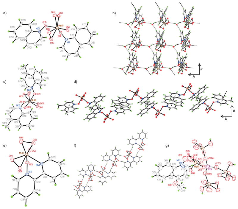
Fig. 3.
(a) Molecular structure of (quinoline-N-oxide)2[Mo(η2-O2)2O] 26 in the crystal. (b) Two-dimensional hydrogen-bonded layer resulting from the interlinking of hydrogen-bonding chains in the crystal of 26·H2O, viewed down the c-axis. (c) Molecular structure of (4-methylquinoline-N-oxide)2[Mo(η2-O2)2O] 27. (d) Extended chains formed by π–π interactions between molecules of 27, viewed down the b-axis. (e) Molecular structure of (κN,κO-2,2′-dipyridyl-N-oxide)[Mo(η2-O2)2O] 28. (f) Hydrogen-bonded chain of centrosymmetric dimers of 28 viewed down the b-axis. (g) X-ray structure of (4-methoxyquinoline-N-oxide)·H3P[OMo(η2-O2)2O]4 29. Thermal ellipsoids are at 50% probability.

