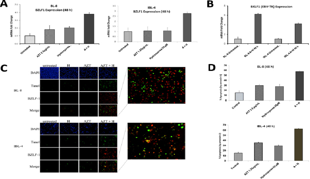
Figure 3.
AZT-induced EBV-lytic reactivation and apoptosis in lymphoma cell lines is potentiated by hydroxyurea (HU). Shown is the synergistic effect of AZT and HU in inducing the EBV-lytic cycle and apoptosis in Burkitt (BL-8) and immunoblastic (IBL-4) lymphoma cell lines at specified times and treatments. (A) Graphs depict relative BZLF1 gene expression by Taqman PCR. Error bars are representative of standard deviation of three separate experiments. (B) Graph depicts relative BXLF1 gene expression by PCR assay in untreated and AZT – HU-treated cell lines. Error bars are representative of standard deviation of three separate experiments. (C) Nuclei stained with DAPI (blue signal), TUNEL (green signal, indicative of fragmented DNA, a marker of apoptosis) and EBV early lytic protein BZLF-1 (red signal) are shown in BL-8 and IBL-4 cells (A, AZT; H, hydroxyurea). Cytospins used for these experiments were prepared from cells treated in (A). Right amplified panels show numerous co-stained TUNEL and BZLF-1 positive cells (red-green or orange color) after treatment with AZT plus hydroxyurea. (D) Graphs depict percentage of apoptosis by Annexin V/PI assays done after 48 h of incubation with control, AZT, HU or their combination. Error bars are representative of standard deviation of three separate experiments.

