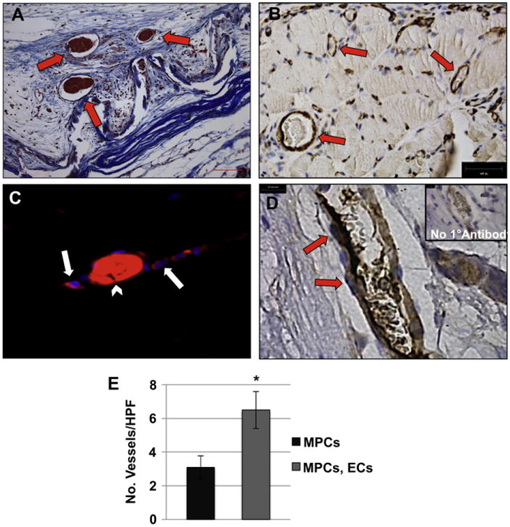
Fig. 2.

Integration of implanted ECs in the TE muscle vasculature. (A) Masson's trichrome stain demonstrated the presence of blood vessels within the scaffold (arrows). The collagen scaffold is stained light blue. Scale bar: 200 μm. (B) Immunohistochemistry for CD146 showed pericyte-covered blood vessels (arrows) distributed within the engineered muscle tissue. (C) Fluorescent microscopy showed integration of DiI-labeled ECs (red, arrow) into a blood vessel containing erythrocytes (autofluorescence in red, arrow-head). (D) The identity of implanted human ECs was confirmed using a human-specific anti-CD31 antibody (arrows, positive CD31 staining). Inset: control staining without primary antibody. (E) Blood vessels were quantified in constructs seeded with MPCs only to those dual seeded with MPCs and ECs, by counting the number of vessels visibly containing erythrocytes from ≥20 random trichrome stained HPFs. Data are expressed as means ± SEM for number of HPFs. Scale bars: 200 μm (A), 10 μm, (BeD). *P < 0.04. (For interpretation of the references to colour in this figure legend, the reader is referred to the web version of this article.)
