
Fig. 4.
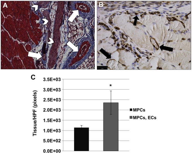
Combination of ECs, together with MPCs, improves muscle tissue formation in vivo. (A) Masson's trichrome stain was used to demonstrate the formation of muscle tissue (red) on the BAM scaffold. Arrows indicate blood vessels within the muscle tissue. Arrowheads indicate nerve bundles. Scale bar: 100 μm (B) CD146 IHC was used to demonstrate the presence of blood vessels localized within the muscle tissue (black arrows). Many small capillary-like vessels were found associated with the myofibers. Scale bar: 50 μm. (C) The amount of tissue was quantitated from ≥10 random trichrome stained HPFs using ImageJ analysis software. The data showed an increase in tissue area when scaffolds were seeded with both ECs and MPCs. Data are expressed as means ± SEM for number of HPFs. *P < 0.03. (For interpretation of the references to colour in this figure legend, the reader is referred to the web version of this article.)