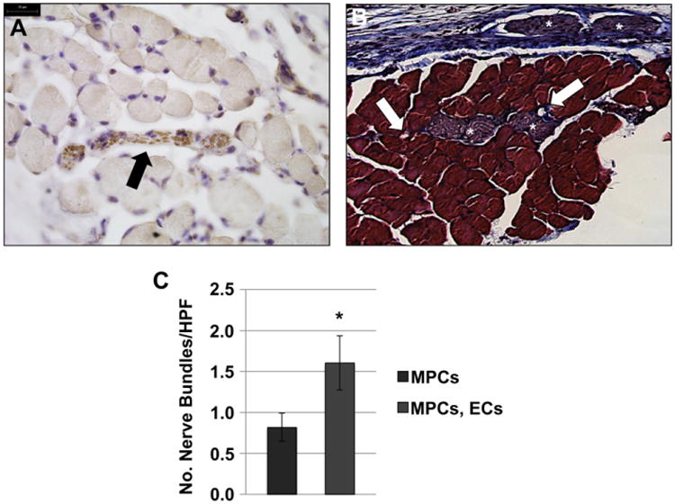
Fig. 6. Combination of ECs with MPCs improves muscle tissue innervation in vivo.

(A) βIII tubulin staining indicated nerve bundles interspersed with the myofibers on the scaffolds. (B) Neuro-vascular bundles were observed in trichrome stained sections of explanted TE muscle constructs (asterisks - nerve bundles; arrows - blood vessels). Scale bars: 25 μm. (C) Quantification of βIII tubulin-positive nerve bundles in ≥20 random HPFs, showed a trend towards an increase of nerve bundle number when scaffolds were seeded with both ECs and MPCs. Data are expressed as means ± SEM for number of HPFs. *P < 0.06.
