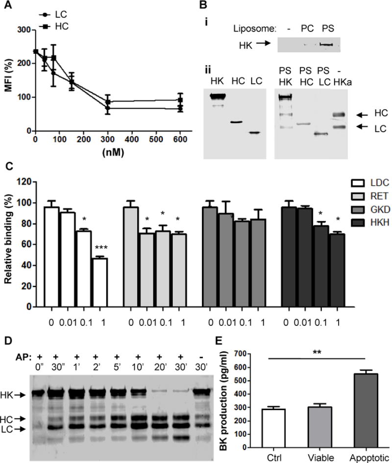
Figure 6. Both of heavy chain and light chain are involved in HK binding to apoptotic cells and PS.

(A) HC and LC inhibit the binding of HK to apoptotic cells. Apoptotic cells were incubated with HC and LC at the indicated concentrations at 4°C for 20minutes, followed by addition of biotin-HK (100 nM) and PE-labeled avidin. The rest steps were as described in the legend for Figure 5(B). Shown are the measurements from 3 separate experiments. (B) HK binds to PS liposome via its heavy chain and light chain. (i) Five μ g of HK protein were incubated with PBS (−), 50 nM PC or PS liposomes in Tyrode’s buffer containing 0.35% BSA and 50 μM ZnCl2 at 4°C for 30 minutes. After ultracentrifugation at 55000 rpm, the pellets were washed twice and solubilized by sample buffer and analyzed by immunoblotting with polyclonal anti-HK antibody. (ii) Before (left panel) or after (left panel) five μg of HK, recombinant heavy chain (HC) or light chain (LC) of HK were incubated with 50 nM PS liposomes. After incubation with PS liposomes, the samples were ultracentrifuged and the pellets were collected. Samples were analyzed by immunoblotting with polyclonal anti-HK antibody. HKa serves as control for molecular weight. (C) In the presence of various peptides at the indicated concentrations, apoptotic cells were incubated with 100 nM biotinylated HK (HK-B), followed by staining with PE-avidin and quantitation by flow cytometry. (D) HK at 30 nM was incubated with apoptotic cells in the presence of prekallikrein (30 nM) at 37°C for the indicated time periods. Apoptotic cells were sonicated and centrifuged at 800 g for 5 minutes. Pellets containing membrane fraction was analyzed by Western blotting using anti-HK Ab. (E) Bradykinin production. Fresh human citrated cell-free plasma was incubated with 5×10⁵ viable cells (Viable), apoptotic cells (Apoptotic) or equal volume of PBS (Ctrl) at 37°C for 30minutes, followed by measurement of bradykinin production. **, p <0.01.
