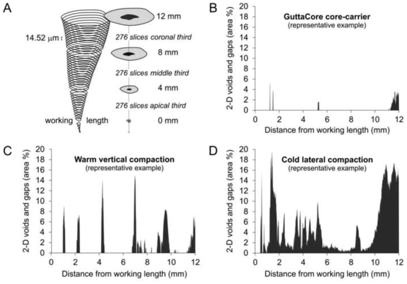
Figure 2.

A. Schematic illustrating the use of axial sections for analyzing the variation in percentage area distribution of gaps and voids along the longitudinal axis of an obturated canal space. At a spatial resolution of 14.52 μm, 276 axial slices were obtained for each of the three canal levels, commencing from the working length (0-4 mm level, 4-8 mm level and 8-12 mm level). Representative examples are shown for the percentage distribution of 2-D gaps and voids in roots obturated using the B. GuttaCore core-carrier technique; C. warm vertical compaction technique; and D. cold lateral compaction technique.
