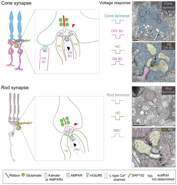
Figure 11. Synaptic connectivity at the OPL.
Schematics and ultrastructure of cone and rod photoreceptor synapses and receptor composition at each synapse type.
ON BC: ON-cone bipolar cell, OFF BC: OFF-cone bipolar cell, RBC: rod bipolar cell, HC: horizontal cell. Metabotropic glutamate receptors (mGluR6) on ON-bipolar cell dendrites mediate a hyperpolarization (sign-inverting) response to glutamate, whereas ionotropic glutamate receptors (AMPA and Kainate receptors) mediate a sign-conserving response in OFF-bipolar cells and horizontal cells. As different species or different OFF-bipolar subtypes express Kainate and/or AMPA receptor both are represented in the schematic, but OFF-bipolar cells in mouse and macaque retina primarily use Kainate receptors for signal transmission through the OPL (see text for details). Red arrow indicates negative feedback and black arrow indicates feedforward modulation. Electron micrographs of rod and cone photoreceptor terminals are from mouse retina. Arrow in electron micrograph points to a ribbon.

