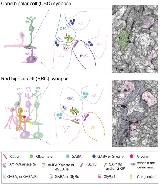
Figure 14. Synaptic connectivity in the IPL.
Schematics showing basic organizations of cone bipolar cell (CBC) and rod bipolar cell (RBC) synapses. Neurotransmitter receptor types are shown. AC: amacrine cell, RGC: retinal ganglion cell. Only AI/A17 and AII amacrine cells are postsynaptic to RBCs. Examples of the ultrastructure of bipolar and amacrine cell synapses in mouse retina are provided in the electron micrographs. Arrow indicates ribbon.

