
FIGURE 8.
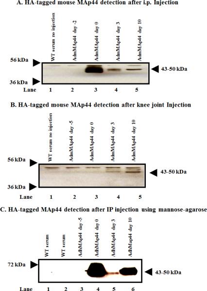
In vivo transduction and expression efficiency of AdmMAp44 or AdhMAp44 assessed by using Western blot analysis for the HA tag on mouse MAp44 in the sera of WT mice before and after the induction of CAIA. Mice were injected in separate studies with AdmMAp44 or AdhMap44 i.p. and also locally in the right knee joint. After SDS-PAGE and transfer to nitrocellulose, the blots were probed with anti-HA rabbit antibody. The presence of a HA band (~43-50 kDa) in serum indicates the successful transduction of cells and protein expression in mice treated with AdmMAp44. A. Presence of HA band in serum at day 0 (lane 3), at day 3 (lane 4) and at day 10 (lane 5) after mice were injected i.p. with AdmMAp44 at day -2 (lane 2). B. Presence of HA band in serum at day 3 (lane 4) and at day 10 (lane 5) after mice were injected in the right knee joint with AdmMAp44 at day -5 (lane 2). Serum from a WT mouse with no injection of adenoviral vectors was used as a negative control (lane 1). C. MAp44 bound to MBL in serum after i.p. injection of AdhMAp44 at day -5. MBL-MAp44 complexes were pulled down using mannose-agarose beads, and the presence of the HA tag on human MAp44 in serum at day 0 (lane 4), day 3 (lane 5), and day 10 (lane 6) was detected using rabbit anti-HA antibody. Serum from a WT mouse without mannose-agarose preparation (lane 1) as well as serum from a WT mouse with no injections with Ad vectors (lane 2) were also examined as controls.