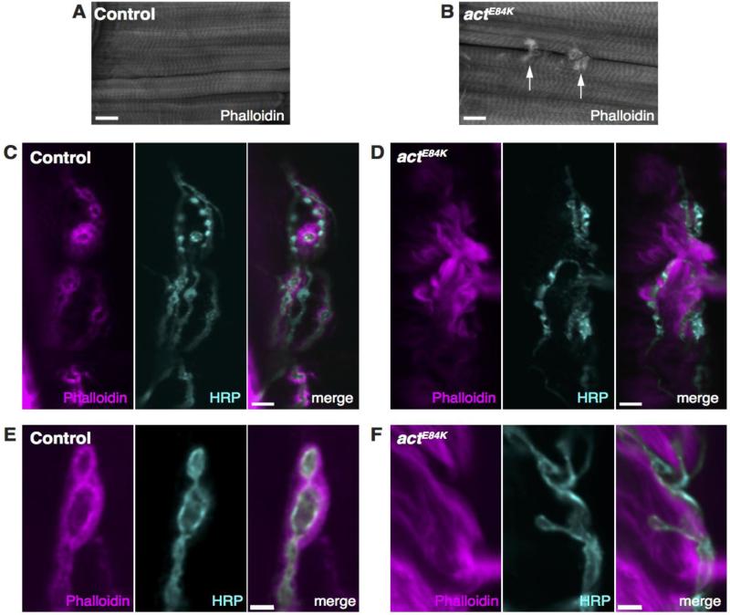
Fig. 4.
The postsynaptic actin cytoskeleton is impaired in actE84K mutants. (A) Normal sarcomere structure observed in control animals revealed by phalloidin staining. (B) Sacromere structure is preserved in actE84K. However, abnormal actin swirls are found that concentrate around the synaptic arbors of the NMJ (arrows). Scale bar in A and B, 20 μm. (C) Phalloidin and anti-HRP staining reveals the normal actin halo surrounding synaptic boutons at control NMJs. (D) The actin halo seen at control NMJs is disrupted in actE84K. Instead, actin forms filamentous swirls that surround boutons. Scale bar in C and D, 10 μm. (E) A high magnification image of the postsynaptic actin cytoskeleton at a control NMJ is shown. (F) The normal halo pattern of actin organization shown in control is lost in actE84K mutants, and wisps of actin filaments are observed instead. Scale bar in E and F, 2 μm.

