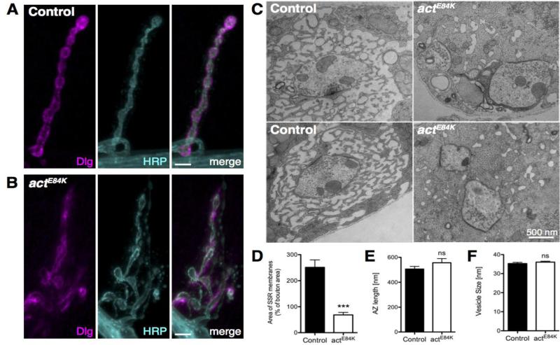
Fig. 6.
Localization of DLG and formation of the subsynaptic reticulum (SSR) is disrupted in actE84K. (A, B) DLG localizes to the SSR in control animals (A), but is severely mislocalized in actE84K mutants (B). Scale bar, 5 μm. (C) Ultrastructural EM analysis shows severe disruption of the SSR in actE84K mutants compared to control. Muscle myofibrils can be observed to directly abut presynaptic membrane (arrowhead) upon loss of SSR membrane folds in actE84K. Scale bar, 500 nm. (D) SSR area is severely reduced in actE84K mutants (N=21 boutons from 3 larvae) compared to control (N=39 boutons from 5 larvae). (E) AZ length is not altered in actE84K mutants (N= 31) compared to control (N=58). (F) Synaptic vesicle diameter is not altered in actE84K mutants (N= 80) compared to control (N=80). Mean and SEM are indicated.

