Figure 2.
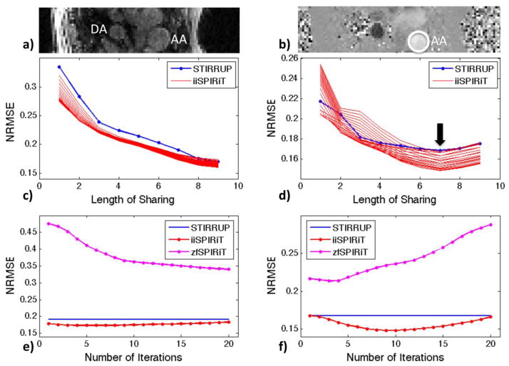
Cross-sectional view of the Ascending Aorta (AA) and Descending Aorta (DA) for: a) Magnitude image, and b) velocity image. Normalized root-mean square errors (NRMSEs) versus length of sharing for: c) magnitude images; and d) through-plane mean velocities within AA. The group of curves with iiSPIRiT (c&d) corresponds to different numbers of iterations (1 to 20). NRMSEs versus number of SPIRiT iterations for: e) magnitude images; and f) through-plane mean velocities within AA. The identified optimal view sharing length of L=7 (15 time frames) was used in e&f.

