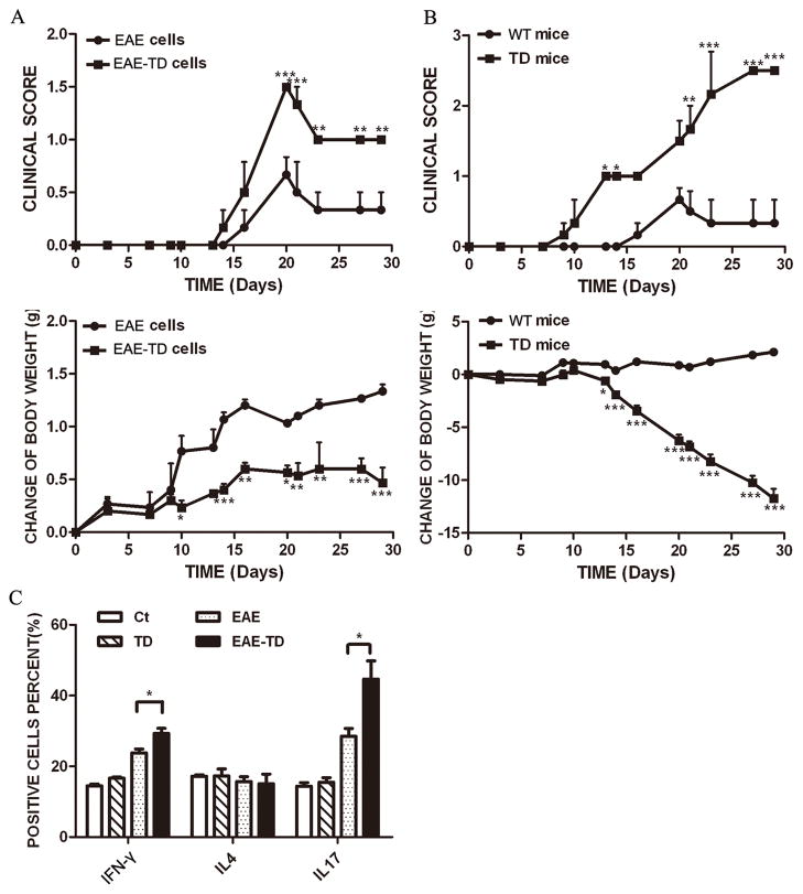
Figure 5. Effect of TD on adoptive EAE model.
A: EAE was induced in C57BL/6 mice by injection of MOG35-55 with or without TD. Encephalitogenic cells were collected from these mice as described under the Materials and Methods and injected into recipient mice (wild-type). The recipient mice were irradiated sublethally (500 rad) 16 hours before the injection. The clinical scores for EAE and body weight were determined as described under the Materials and Methods. B: EAE was induced in C57BL/6 mice by injection of MOG35-55. The encephalitogenic cells were prepared from EAE mice and injected into recipient mice with/without TD. The clinical scores for EAE and body weight were determined as described under the Materials and Methods. Data were presented as mean ± SD; *p<0.05, **p<0.01, ***p<0.001, compared with control group. n = 8 for each group. C: Lymph node cells were isolated from EAE mice with or without TD after 10 days of induction. The percentage of IFN-γ, IL-4 and IL-17 producing cells were measured by FACS. Data were presented as mean ± SD; *p<0.05, compared with EAE group. n = 4 for each group.

