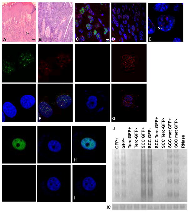
Fig. 1.
Terc−/− mice develop primary epidermal SCC. Tissue sections of primary SCC in Terc+/+ (A) and Terc−/− (B) mice are stained with hematoxylin and eosin. Arrows indicate tumor cells. Stem cells in Terc−/− mice exhibit telomeric DNA damage response and apoptosis. Localization of 53BP1 (FITC) at telomeres (Cy3) in tissue sections Terc−/− (C) and Terc+/+ (D) SCC is shown. Representative sections from three independent experiments are shown. Scale bar = 5 μm. (E) Enlarged photomicrograph showing localization of 53BP1 at telomeres (arrow) in Terc−/− SCC. Localization of 53BP1 protein at telomeres (yellow signals) in sorted stem cells from Terc−/− (F) and Terc+/+ (G) SCC. Apoptosis of sorted stem cells from Terc−/− (H) and Terc+/+ (I) SCC is shown by TUNEL analysis. (J) Telomerase activity in GFP+ and GFP- cells from normal epidermis, Terc negative epidermis, Terc+ and Terc- SCC, and metastatic SCC is shown by TRAP assay. RNase treated extract was used as the negative control.

