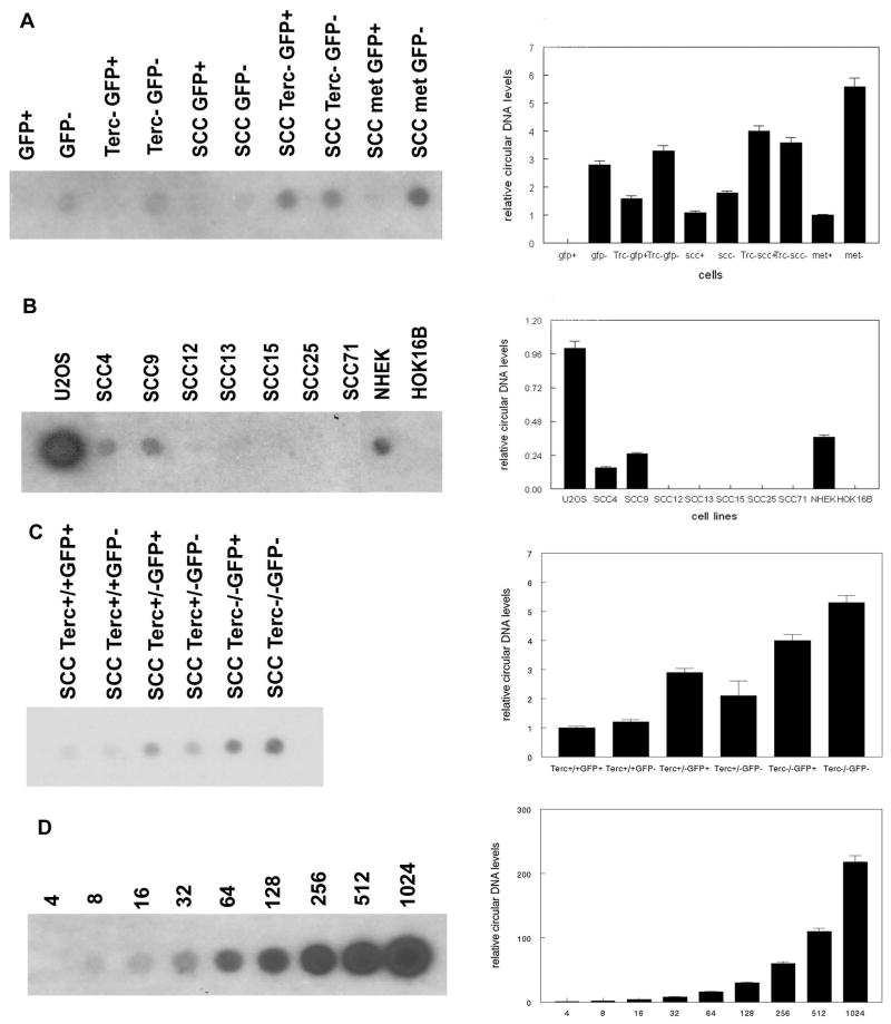
Fig. 4.
Terc+/+ keratinocytes and SCC exhibit telomeric circular DNA. (A) Telomeric circular DNA was amplified from mouse sorted Terc+/+ and Terc−/− epidermal stem (GFP+) and basal cells (GFP−), Terc+/+ and Terc−/− SCC cells, and Terc+/+ metastatic SCC cells and detected by southern blot. (B) Telomeric circular DNA was amplified from human U2OS, SCC, NHEK, and HOK16B cells. (C) Telomeric circular DNA was amplified in GFP+ and GFP− cells from Terc+/+, Terc+/−, and Terc−/− SCC. (D) Control amplifications of synthesized 96mer telomeric circular DNA. Nanograms of circular DNA in each reaction are shown. A minimum of three independent experiments was performed. Quantitation of blots is shown at right.

