Fig. 6.
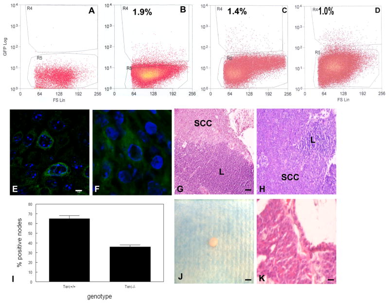
Stem cells from epidermal SCC are tumorigenic and contribute to primary and metastatic tumors. (A) Dissociated epidermal keratinocytes from control mice (A), K15/GFP/Terc+/+ mice (B), K15/GFP/Terc−/− mice (C), and K15/GFP epidermal SCC (D) were subjected to flow cytometric sorting. K15/GFP+ tumor cells were localized by immunofluorescence microscopy in primary (E) and metastatic (F) epidermal SCC. Scale bar = 2 μm. Metastatic SCC in Terc+/+ (G) and Terc−/− (H) mice is shown by hematoxylin and eosin staining. (I) Reduced metastatic SCC in Terc−/− compared to Terc+/+ SCC. (J) A tumor from sorted K15+ epidermal SCC cells subcutaneously injected into nude mice is shown. Scale bar = 5 mm. (K) A tissue section from tumor shown in (J) revealed poorly differentiated SCC. Scale bar = 50 μm.

