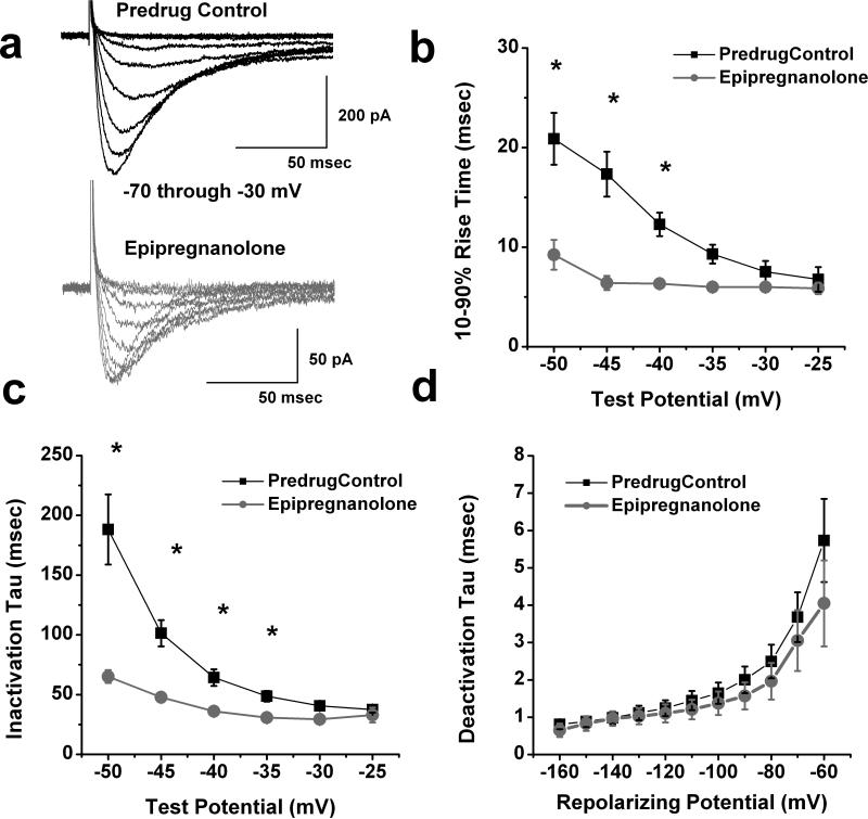
Figure 2. Effects of epipregnanolone of macroscopic T-current kinetics and deactivation in rat DRG cells.
a: Traces represent families of T-currents evoked in a representative DRG cell in predrug control conditions (black traces on top panel) and during application of 10 μM epipregnanolone (gray traces on lower panel) by voltage steps from Vh of -90 mV to Vt from −70 through −30 mV in 5-mV increments. Bars indicate calibration. b,c: We measured time-dependent activation (10%-90% rise time, panel b) and inactivation τ(single exponential fit of decaying portion of the current waveforms, panel c) in 8 DRG cells over the range of test potentials from −50 mV to −25 mV before (■) and after application of 10 μM epipregnanolone (●). Note that epipregnanolone speeded T-current kinetics at more negative Vt. Symbol * indicates significance of p < 0.05.
d: Deactivating tail currents in control predrug conditions (■) and after application of 10 μM epipregnanolone (●) were fit with a single exponential function. The resulting tau values are plotted (n = 6). All points are not statistically significant between two groups (p > 0.05).

