
Figure 1.
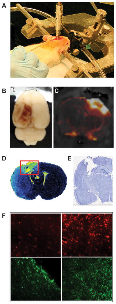
Acute and chronic pathology resulting from experimental traumatic brain injury. A. Representation of the controlled cortical impact (CCI), one of the most widely used models of experimental TBI. The injury is administered directly to the brain of an anesthetized animal with a computer-controlled, air-driven piston. This injury results in breakdown of the BBB and the extravasation of blood components into the brain parenchyma that can be observed through gross evaluation (B) and gadolinium-enhanced magnetic resonance imaging (C). A robust immune response results over a period of weeks (D) observed through binding of [3H]-PK11195 to the translocator protein (18 kDa) expressed on the outer mitochondrial membrane of activated microglia) that ultimately leads to tissue loss and cavity formation (E). (F) Further evidence of this response can be observed through immunohistochemistry. The uninjured hemisphere (left column) has relatively low expression of microglia/macrophage marker ED1 (top row) or astrocyte marker GFAP (bottom row) compared to the lesion site (right column).