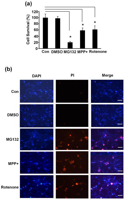
Fig. 4.
Impairment of neuronal viability following disruption of proteasome degradation pathway. (a) SN4741 cells were treated with MG132 (2.5 μM), MPP+ (25 μM) or rotenone (100 nM) treatment for 24 hours. Cell viability was measured by WST-1 assay. The experiments were repeated 4 times. (b) SN4741 cells were treated as in (a) and than stained with PI (Prodidium Iodide) and DAPI. The experiments were repeated 4 times. Bar=200 μm. *, p<0.01.

