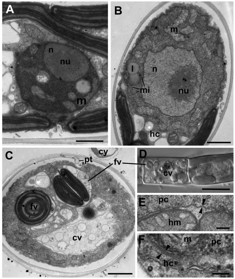
Figure 4. Structure of the Aphelidium aff. melosirae young trophont seen on ultrathin sections under TEM (A-C, E-F) and LM (D).
A – one of the first stages of trophont development after invasion, B – bigger uninuclear trophont adpresses the host cytoplasm, C – trophont with two food vacuoles and central vacuole, D – LM of the same stage as in C. E – host plasma membrane and food vacuole membrane (arrowheads) delimiting food vacuole. F – two membranes (arrowheads), bordering the parasitoid cell in the host. Abbreviations: cv-central vacuole, cw-cyst wall, cy-cyst, er-endoplasmic reticulum, fv-food vacuoles, hc-host cytoplasm, hm-host mitochondrion, hw-host cell wall, l-lipid globule, m-mitochondrion, mi-microbody, n-nucleus, nu-nucleolus, pc-parasitoid cytoplasm, pt-penetration tube. Scale bars: A-C – 2 μm, D – 10 μm, E – 400 nm, F – 100 nm.

