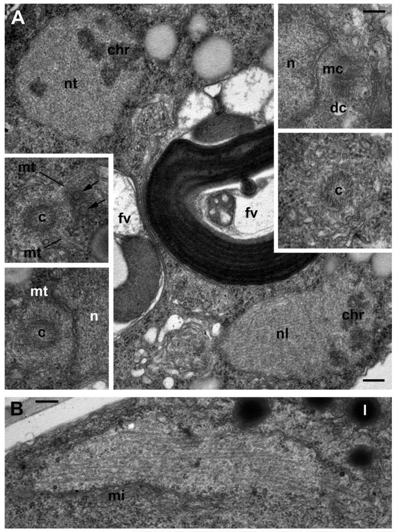
Figure 5. Centrosome structure and nuclear division in the young trophont of Aphelidium aff. melosirae.
A – prometaphase in two dividing nuclei with transverse (nt) and nearly longitudinal (nl) sections of mitotic spindle. Inserts show centrioles (c) with radiating microtubules (mt), centrosome composed of two orthogonal centrioles: mother (mc) and daughter (dc). Arrows show nuclear pores. B – elongated nucleus at late anaphase/early telophase. Other abbreviations as in previous Figures. Scale bars: A – 500 nm, A inserts – 200 nm, B – 250 nm.

