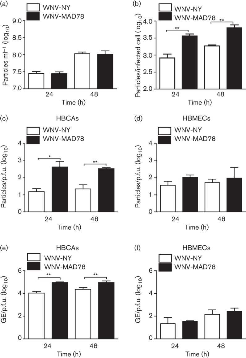
Fig. 2.

Infectivity of HBCA-derived particles. HBCAs were infected with WNV-NY or WNV-MAD78 (m.o.i. 0.01). (a) Total viral particle production at 24 and 48 h post-infection. Culture supernatants were removed at the indicated times and particle levels quantified using the ViroCyt virus counter. Values represent the mean±se number of particles ml–1 of three independent experiments. (b) Particle production per infected cell was determined by dividing the total number of viral particles present in the culture supernatant as determined in (a) by the total number of infected cells as determined by flow cytometry. Values represent mean±se of three independent experiments. **P<0.01. (c–f) Infectivity of astrocyte-derived (c, e) and HBMEC-derived (d, f) WNV particles. (c, d) The concentration of total virus particles and infectious particles was determined using the ViroCyt virus counter and plaque assays on Vero cells, respectively. Values represent the mean±se ratio of total particles compared with infectious particles from three independent experiments. *P<0.05, **P<0.01. (e, f) The concentration of viral genomes (GE) and infectious particles was determined using qRT-PCR and plaque assays on Vero cells, respectively. Values represent the mean±se ratio of viral genomes compared with infectious particles from three independent experiments. **P<0.01.
