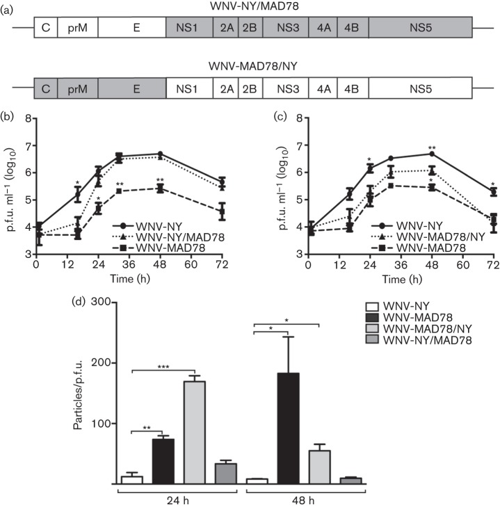
Fig. 6.
Replication of WNV-NY/MAD78 and WNV-MAD78/NY in HBCAs. (a) Schematic representation of WNV-NY/MAD78 and WNV-MAD78/NY. Regions derived from WNV-NY are in white and regions from WNV-MAD78 are in grey. (b–d) HBCAs were infected with WNV (m.o.i. 0.01). (b, c) Growth curves of WNV-NY, WNV-MAD78 and WNV-NY/MAD78 (b) or WNV-MAD78/NY (c). Culture supernatants were collected at the indicated times and viral titres determined by plaque assay on Vero cells. Values represent the mean±se p.f.u. ml–1 of at least three independent experiments. Statistical significance comparing replication of both chimeras in relation to WNV-NY (indicated above the line representing WNV-NY) and WNV-MAD78 (indicated above the line representing WNV-MAD78) is shown. *P<0.05, **P<0.01. (d). Infectivity of astrocyte-derived WNV-NY, WNV-MAD78, WNV-MAD78/NY and WNV-NY/MAD78 particles. The concentration of total virus particles and infectious particles (b, c) was determined using the ViroCyt virus counter and plaque assays on Vero cells, respectively. Values represent the mean±se ratio of total particles to infectious particles from three independent experiments. *P<0.05, **P<0.01, ***P<0.005.

