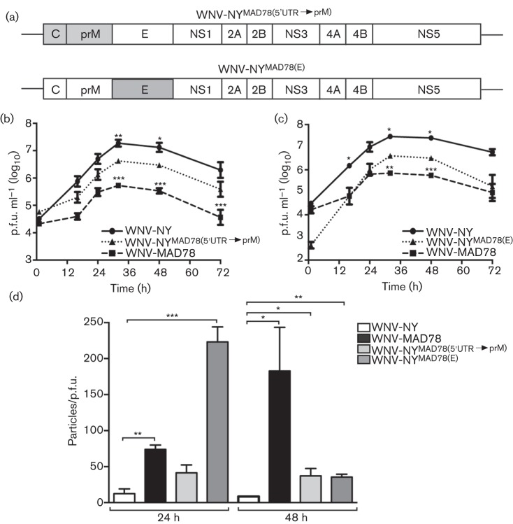
Fig. 7.
Replication of WNV-NYMAD78(5′UTR→prM) and WNV-NYMAD78(E) in HBCAs. (a) Schematic representation of WNV-NYMAD78(5′UTR→prM) and WNV-NYMAD78(E). Regions derived from WNV-NY are in white and regions from WNV-MAD78 are in grey. (b–d) HBCAs were infected with the indicated viruses (m.o.i. 0.01). (b, c) Growth curves of WNV-NY, WNV-MAD78 and WNV-WNV-NYMAD78(5′UTR→prM) (b) or WNV-NYMAD78(E) (c). Culture supernatants were collected at the indicated times and viral titres determined by plaque assay on Vero cells. Values represent the mean±se p.f.u. ml–1 of at least three independent experiments. Statistical significance comparing replication of both chimeras in relation to WNV-NY (indicated above the line representing WNV-NY) and WNV-MAD78 (indicated above the line representing WNV-MAD78) is shown. *P<0.05, **P<0.01, ***P<0.005. (d) Infectivity of astrocyte-derived WNV-NY, WNV-MAD78, WNV-NYMAD78(5′UTR→prM) and WNV-NYMAD78(E) particles. The concentration of total virus particles and infectious particles (b, c) was determined using the ViroCyt virus counter and plaque assays on Vero cells, respectively. Values represent the mean±se ratio of total particles to infectious particles from three independent experiments. *P<0.05, **P<0.01 ***P<0.005.

