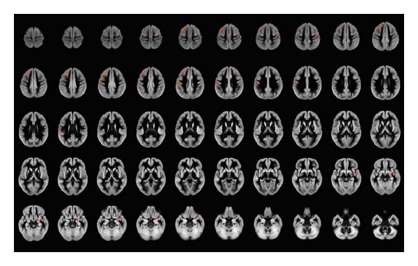
Figure 4.

Areas of significant gray matter differences at ANCOVA (including correction for age and total intracranial volume, less than 1 false-positive cluster expected, P < 0.005) between responder and nonresponder schizophrenia patients, superimposed onto the average of the normalized GM volumes. Right side of the brain is at the observer's right. Clusters of significantly reduced GM volume in nonresponder schizophrenia patients compared to responder schizophrenia patients are detected mainly the frontal lobes bilaterally, more extended on the left where the pre- and postcentral gyri are involved, while on the right mainly middle frontal gyrus and insula are involved.
