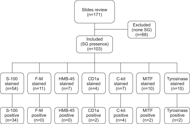
Fig. 1.
We retrospectively reviewed 171 H&E slides taken from the scalp and facial skin. Among them, 103 slides containing sebaceous gland (SG) tissues were finally included in the study. Of these, 54 were stained with the antiserum to S-100, 11 with Fontana-Masson (F-M) stain, 7 with antiserum to human melanoma black-45 (HMB-45), 4 with antiserum to CD1a, 7 with antiserum to c-kit, 10 with antiserum to microphthalmia-associated transcription factor (MITF), and 10 with antiserum to tyrosinase. Out of the 54 specimens of human SGs, 34 slides showed S-100-positive cells. All specimens showed negative staining for F-M (11 specimens) and HMB-45 (7 specimens). In addition, 2 specimens were found to be CD1a-positive, and 4 were c-kit-positive. Out of 10 specimens of human SGs, 2 showed MITF-positive cells, and 2 others showed positive staining for tyrosinase.

