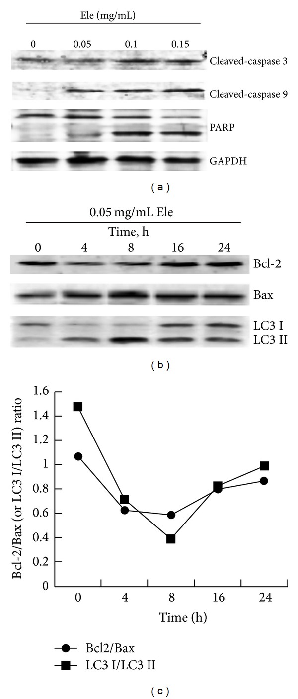
Figure 3.

Ele-induced apoptosis and autophagy of HepG2 cells. HepG2 cells were untreated or treated with different concentration of Ele for 24 h or with 0.05 mg/mL Ele for different times. Apoptosis-related proteins (a) and autophagy-related proteins (b) were detected by Western blotting. The Bcl-2/Bax protein ratio and LC3 I/LC3 II protein ratio were shown in (c).
