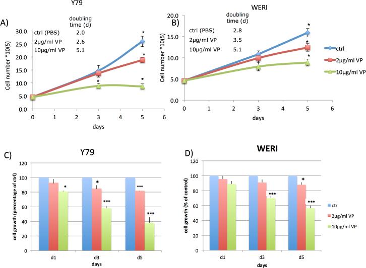
Figure 1. Verteporfin (VP) inhibits growth of retinoblastoma cells Y79 and WERI without light activation.
Y79 and WERI retinoblastoma cells were left untreated (PBS control) or treated with VP for five days; control (blue), VP concentration 2μg/ml (red) and 10μg/ml (green).
(A,B) VP treatment resulted in an inhibition of cell growth and a decrease of the cell number of Y79 and WERI cells in a dose-dependent manner. The doubling time was increased.
(C,D) VP treatment resulted in a significant, time- and dose-dependent inhibition of Y79 and WERI cell growth and viability as determined by MTT assays. The results are expressed as percentage of growth (%) relative to control values. Results are average of three independent experiments. Data are presented as mean +/− SEM (n=9, *p<0.05, *** p<0.001).

